



When a dentist recommends X-rays, many patients have two concerns: what the images might reveal and how much radiation they will receive.
You may also hear that an office uses “digital X-rays” and wonder whether that is meaningfully different from the film images you remember biting down on years ago.
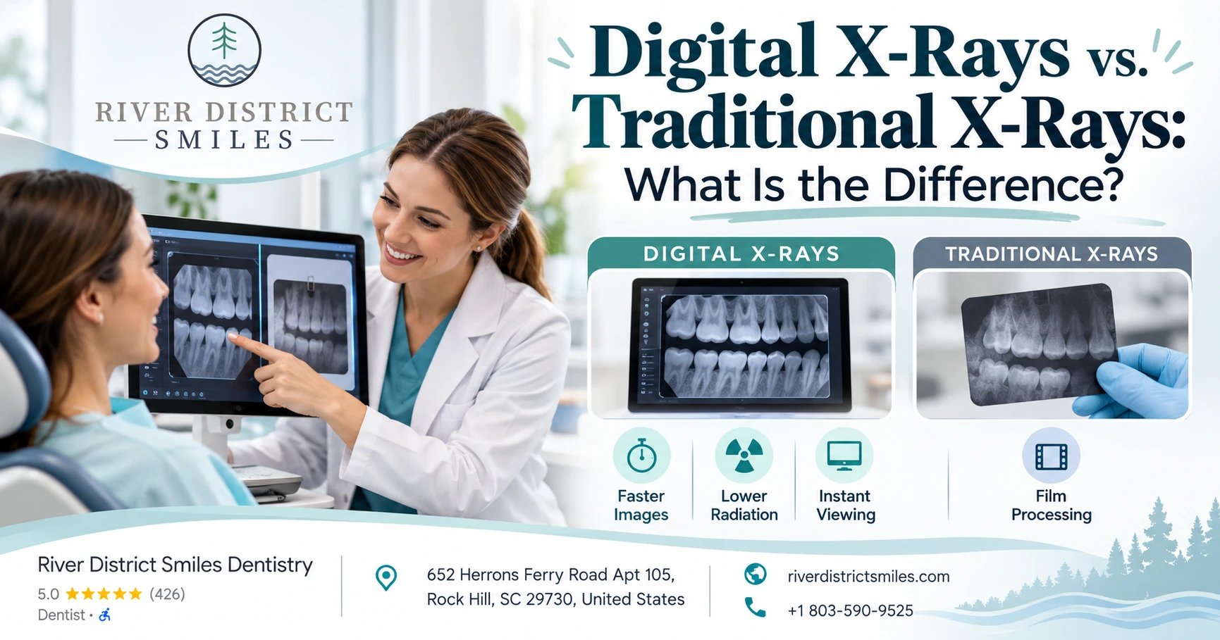
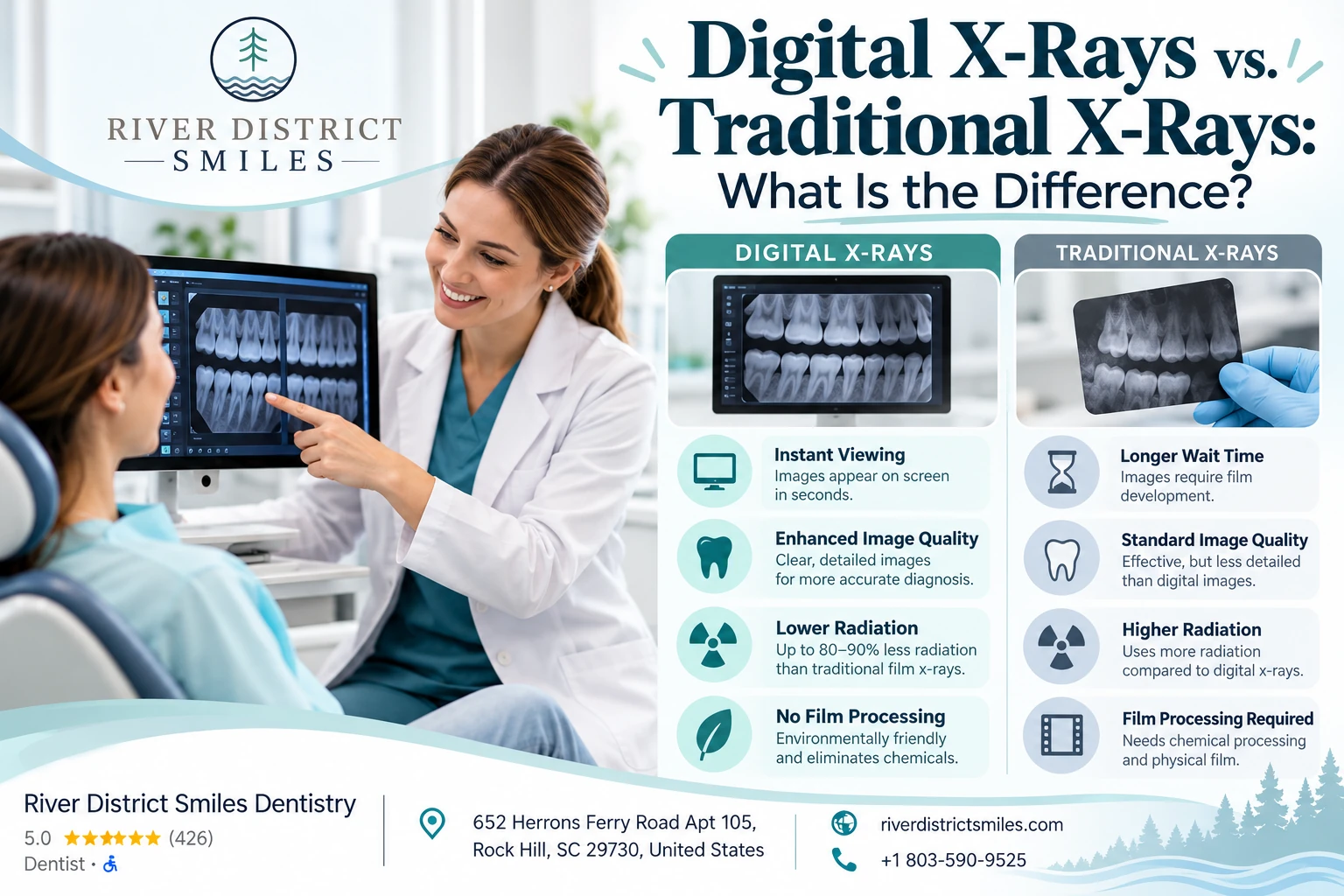
Digital and traditional dental X-rays use the same basic form of radiation to create an image. The difference is how that image is captured, processed, viewed, and stored. Digital X-rays usually produce images faster, allow easier enlargement and sharing, and generally require less radiation than conventional film.
Traditional film can still produce useful diagnostic images. However, digital imaging has become the more practical choice in many modern dental offices.
Digital vs. traditional X-rays at a glance
| Digital dental X-rays | Traditional film X-rays |
| Image is captured by an electronic sensor or imaging plate | Image is captured on dental film |
| Appears on a computer within seconds or minutes | Must be chemically developed |
| Can be enlarged and adjusted for closer review | Physical film has fewer viewing options |
| Usually requires less radiation than conventional film | Radiation needs depend partly on film speed |
| Easy to store, compare, and securely transfer | Physical films require storage or scanning |
| No chemical processing | Requires developing chemicals |
| Sensor may feel thicker for some patients | Film packet may feel thinner or more flexible |
How do dental X-rays work?
Both digital and traditional X-rays use a controlled beam of ionizing radiation.
The beam passes through the teeth and surrounding structures before reaching a receptor. Dense materials, such as enamel and metal restorations, absorb more of the beam and appear lighter. Less-dense areas allow more radiation through and appear darker.
This contrast helps dentists identify conditions that cannot always be seen during a visual examination. The FDA explains that radiography creates a static image by passing an X-ray beam through the body and recording the pattern that reaches a detector.
Dental X-rays may help reveal:
- Cavities between teeth
- Decay beneath some fillings or crowns
- Bone loss around teeth
- Infection near a tooth root
- Impacted or unerupted teeth
- Certain cysts or other abnormalities
- Developing permanent teeth
- Implant position and available bone
- Problems that may affect a root canal or extraction
An X-ray does not provide every answer by itself. It must be interpreted along with your symptoms, dental history, and clinical examination.
What is a digital dental X-ray?
A digital X-ray records the image electronically rather than on film.
For an image taken inside the mouth, the dental team may place either a wired sensor or a thin phosphor imaging plate beside the teeth. The X-ray exposure is made in much the same way as with film.
With a direct digital sensor, the image usually appears on the computer almost immediately. A phosphor plate must first be passed through a scanner, but it still avoids chemical film development.
Digital images can then be:
- Enlarged
- Viewed with different contrast or brightness settings
- Compared with previous images
- Displayed while the dentist explains a finding
- Stored in the electronic patient record
- Securely transferred to another dental provider
The ADA’s current radiation-safety recommendations favor digital receptors over conventional film as part of reducing exposure while maintaining diagnostically useful images.
What is a traditional dental X-ray?
A traditional dental X-ray uses a small packet of film as the image receptor.
After exposure, the film must be developed using chemicals or an automated processing system. Once processed, the image can be viewed on an illuminated screen or mounted with other dental films.
Traditional film is capable of producing diagnostically acceptable images when it is exposed, developed, stored, and viewed correctly. Earlier ADA and FDA guidance recognized both film and digital systems as suitable diagnostic methods.
The limitations are mostly practical:
- Processing takes longer
- Developing errors can affect image quality
- Physical films can be scratched, faded, misplaced, or damaged
- Enlarging a small area is more difficult
- Copying and transferring images takes additional steps
- Chemical processing creates waste and requires quality control
Do digital X-rays use less radiation?
Generally, yes.
Digital receptors are more sensitive than many conventional films, so they can usually produce a useful image with a shorter exposure. River District Smiles states that its digital imaging system uses significantly less radiation than traditional film while producing images that can be reviewed immediately.
The exact difference is not the same in every office. Radiation exposure also depends on:
- The type of sensor or film
- The X-ray equipment
- Exposure settings
- The size of the area being imaged
- The patient’s size and age
- How well the equipment is maintained
- Whether the image must be retaken
- The use of beam-limiting technology
Faster conventional film can also reduce exposure compared with older, slower film. The FDA has encouraged dental professionals who still use film to use faster film speeds to lower the dose required.
The most important safety issue is not simply whether an office uses film or digital technology. It is whether the dentist orders only the images needed and uses appropriate equipment and technique.
Are digital dental X-rays safe?
Dental X-rays involve a low level of radiation, but they are not completely radiation-free.
The dentist should weigh the diagnostic benefit against the exposure and order imaging only when it is likely to affect diagnosis or care. The FDA advises that dental radiographs should be selected according to professional judgment and the individual patient’s needs rather than taken automatically.
Current ADA recommendations emphasize:
- Ordering only clinically necessary images
- Using previous images when they remain useful
- Using digital receptors rather than conventional film when possible
- Limiting the beam to the area that needs evaluation
- Positioning the patient carefully to avoid retakes
- Using three-dimensional imaging only when lower-exposure images will not provide the information needed
For most patients, the diagnostic benefit of a necessary dental X-ray outweighs the small radiation risk.
Why does the dentist need X-rays if nothing hurts?
Many dental problems do not cause symptoms in their early stages.
A cavity between two teeth may not be visible during the examination. Gum disease can cause bone loss without producing pain. Infection around a tooth root may also exist before noticeable swelling develops.
X-rays can allow a dentist to identify a smaller problem before it becomes:
- A larger cavity
- A broken tooth
- A dental abscess
- A need for root canal treatment
- Advanced bone loss
- A more complicated extraction
That does not mean every patient needs the same set of images at every checkup. Imaging frequency should be based on the patient’s oral health, age, dental development, symptoms, treatment history, and risk of new disease.
Are digital images clearer than film?
Digital images are often easier to evaluate because the dentist can enlarge them and adjust the display.
For example, the dentist may zoom in on:
- A suspicious area between teeth
- The edge of a filling or crown
- Bone surrounding an implant
- The tip of a tooth root
- A possible fracture
- Changes between current and previous images
Image-processing tools can help the dentist see certain details, but they do not create information that was not captured in the original exposure. A poorly positioned or blurred digital image may still need to be retaken.
Traditional film can also provide excellent detail when it is processed correctly. Digital systems mainly improve speed, consistency, viewing flexibility, and communication.
Are digital X-rays more comfortable?
The answer depends on the type of digital receptor.
A wired digital sensor may feel thicker and more rigid than a traditional film packet. This can be uncomfortable for patients who have:
- A small mouth
- A sensitive gag reflex
- A shallow palate
- Tender gums
- Large bony areas inside the lower jaw
- Difficulty opening widely
Phosphor plates tend to feel thinner and more flexible, making them more similar to film.
Comfort can often be improved by:
- Using a smaller sensor
- Repositioning the receptor
- Allowing short breaks
- Having the patient breathe slowly through the nose
- Taking only the necessary images
- Using extraoral imaging when clinically appropriate
Tell the dental team if previous X-rays have caused gagging or discomfort. A rushed or painful technique is more likely to lead to movement and retakes.
Do digital X-rays show cavities better?
Digital X-rays can be very useful for detecting cavities, but they do not automatically identify every area of decay.
The dentist combines the images with a visual and tactile examination. Very early enamel changes may be monitored rather than immediately treated, while deeper areas may require a filling or another restoration.
X-rays are especially useful for detecting cavities:
- Between neighboring teeth
- Beneath certain existing restorations
- Along crown margins
- In areas that cannot be seen directly
An image can sometimes make a cavity appear smaller than it actually is because an X-ray is a two-dimensional view of a three-dimensional tooth. The dentist may discover more extensive damage after the area is opened.
What types of digital dental X-rays are there?
“Digital X-ray” can refer to several types of images.
Bitewing X-rays
Bitewings usually show the crowns of the upper and lower back teeth and part of the supporting bone.
They are commonly used to look for:
- Cavities between teeth
- Changes beneath fillings or crowns
- Bone-level changes
- Tartar below the gumline
Periapical X-rays
A periapical image shows the entire tooth, including the root and surrounding bone.
It may be used to evaluate:
- Tooth pain
- Infection
- Root canal treatment
- Trauma
- Root shape
- Bone around an implant
- A tooth before extraction
Panoramic X-rays
A panoramic image is taken by a machine that rotates around the head. It provides a broad view of the teeth, jaws, jaw joints, and surrounding structures.
It may be useful for:
- Wisdom teeth
- Missing or developing teeth
- Jaw conditions
- General surgical planning
- Certain orthodontic evaluations
A panoramic image does not replace detailed bitewings or periapical images when close evaluation of cavities or individual roots is needed.
Cone-beam CT imaging
Cone-beam computed tomography, or CBCT, produces a three-dimensional view.
It may be used for:
- Dental implant planning
- Complicated root canal anatomy
- Impacted teeth
- Selected jaw conditions
- Surgical planning
- Certain fractures or infections
A CBCT scan usually exposes the patient to more radiation than a limited two-dimensional dental image, so it should be used only when the additional three-dimensional information is expected to change diagnosis or treatment. The FDA and ADA both emphasize appropriate patient selection for CBCT.
Does digital mean three-dimensional?
No.
Most routine digital dental X-rays are still two-dimensional. The word “digital” describes how the image is captured and stored.
A digital bitewing and a film bitewing show the same general type of two-dimensional view. The digital version simply uses an electronic receptor rather than conventional film.
A cone-beam CT scan is a different imaging method that produces three-dimensional information. It is not necessary for every dental examination.
How often should dental X-rays be taken?
There is no one schedule that is right for every patient.
A person with a history of frequent cavities may need bitewing images more often than someone who has had no recent decay and has very low risk. A child with developing teeth may have different needs than an adult with a stable mouth.
The decision may depend on:
- Whether you are a new or established patient
- Your cavity history
- Gum health
- Age and stage of dental development
- Existing crowns, bridges, or implants
- Current pain or swelling
- Previous root canal treatment
- Planned dental procedures
- Whether useful recent images are available
The ADA’s 2026 patient-selection recommendations support choosing imaging according to the clinical situation rather than using a fixed routine for every patient.
Ask why an image is being recommended and what the dentist expects to learn from it. A clear answer should relate the X-ray to your health history, symptoms, or treatment plan.
Can my new dentist use X-rays from another office?
Often, yes.
Previous images may help the new dentist:
- Compare changes over time
- Avoid unnecessary repeat imaging
- Review earlier treatment
- Understand the history of a tooth
- Plan which new images are actually needed
However, additional images may still be recommended when the previous ones are:
- Too old for the current concern
- Blurred or incomplete
- Taken at the wrong angle
- Missing the area being evaluated
- Difficult to open or transfer
- Not appropriate for the planned procedure
Current ADA safety guidance encourages dental professionals to use previously acquired images when they remain diagnostically useful.
You can ask your previous office to send the original digital files rather than screenshots or printed copies, since the originals usually retain more detail.
Do you need a lead apron for digital dental X-rays?
Practices around shielding have changed as imaging equipment and safety guidance have improved.
Recent ADA recommendations state that routine thyroid and abdominal shielding is no longer necessary during dental imaging when modern equipment, proper beam limitation, patient positioning, and appropriate technique are used.
Some dental offices may continue to use protective aprons because of state requirements, office policy, equipment considerations, or patient preference.
The absence of a lead apron does not by itself mean the X-ray is being performed unsafely. Modern safety places greater emphasis on:
- Ordering only necessary images
- Limiting the beam
- Using appropriate exposure settings
- Avoiding retakes
- Maintaining and testing equipment
- Selecting the correct imaging method
Patients who are concerned should feel comfortable asking the dental team to explain the office’s current protocol.
Can you have dental X-rays during pregnancy?
Tell the dental team if you are pregnant or may be pregnant.
Necessary dental imaging can still be performed when it is important for diagnosis or urgent treatment. Delaying care for a serious infection can create its own health risks.
The dentist may postpone elective imaging that is unlikely to affect immediate care, but urgent symptoms such as pain, swelling, trauma, or infection may require an X-ray to make a safe decision.
The recommendation should be based on the reason for the image—not simply on whether the system is digital or film.
Are dental X-rays safe for children?
Children should receive dental X-rays only when the images are expected to provide useful information.
They may need imaging to evaluate:
- Cavities between teeth
- Tooth development
- Missing or extra teeth
- Dental injuries
- Eruption problems
- Orthodontic concerns
- Infection
Because children are smaller and their teeth and jaws are developing, exposure settings and image size should be adjusted appropriately. Current guidance emphasizes patient-specific selection and limiting the imaged area to what is necessary.
A child should not receive a full set of X-rays simply because every other child receives one at the same age.
Are digital X-rays more expensive?
The patient fee is often based on the type and number of images rather than whether the office uses digital or film equipment.
Costs may vary according to whether the dentist takes:
- One focused X-ray
- A set of bitewings
- A panoramic image
- A full-mouth series
- A cone-beam CT scan
Digital systems are expensive for a practice to purchase and maintain, but they also remove film and chemical-processing costs. As a result, digital X-rays are not automatically more expensive for the patient.
Many dental insurance plans cover routine diagnostic images according to frequency limits. Coverage varies based on the plan, the type of image, and whether it is considered necessary.
Before imaging, you can ask:
- Which X-rays are being recommended?
- Why are they needed?
- What will they cost?
- Does insurance have a frequency limit?
- Are usable images available from my previous dentist?
- Is a three-dimensional scan included or charged separately?
What are the disadvantages of digital X-rays?
Digital imaging offers meaningful advantages, but it is not perfect.
Possible limitations include:
- Thick or rigid sensors may be uncomfortable
- Equipment and software can malfunction
- Poorly positioned images still require retakes
- Image enhancement can be overused or misinterpreted
- Digital records require secure storage and proper backups
- Not every office system communicates easily with another
- A two-dimensional digital image still has the limitations of any two-dimensional view
Technology improves the process, but accurate diagnosis still depends on proper technique and clinical judgment.
Are traditional X-rays unsafe?
No.
Traditional dental film is not automatically unsafe. It can produce useful images at an appropriately controlled dose, particularly when faster film and proper technique are used.
The main issue is that modern digital receptors can generally achieve the same diagnostic goal more efficiently and with lower exposure. That is why current safety recommendations favor digital imaging.
The age of the equipment alone does not determine safety. Maintenance, calibration, beam limitation, film speed, processing quality, and patient selection all matter.
When digital X-rays are usually the better choice
Digital imaging is generally preferable when:
- The office wants to minimize radiation exposure
- Images need to be viewed immediately
- The dentist wants to enlarge or compare specific areas
- Records may need to be transferred to a specialist
- The patient benefits from seeing the findings on a screen
- The office wants to avoid chemical film processing
- The dentist needs an efficient record for future comparison
River District Smiles uses digital X-rays and three-dimensional cone-beam imaging as part of its dental technology. The practice states that digital images can be captured quickly and reviewed with patients in real time.
When traditional film may still be used
Film may still be found in:
- Offices transitioning to digital systems
- Settings where digital equipment is unavailable
- Certain educational or institutional environments
- Practices using properly maintained film systems with fast film
The more important question is whether the image is diagnostically useful and whether exposure has been kept as low as reasonably appropriate.
Questions to ask before dental X-rays
You can ask:
- What type of X-ray are you recommending?
- What are you looking for?
- Will the image change today’s treatment decision?
- Do you have recent images from my previous dentist?
- Is this a two-dimensional image or a 3D scan?
- How is the exposure adjusted for my size and needs?
- Why is a CBCT scan necessary?
- What will the images cost?
- Will insurance cover them?
- Can you show me what you see?
A dentist should be able to explain the reason for the image in straightforward language.
The bottom line
Digital and traditional X-rays use the same basic type of radiation, but digital systems capture the image electronically. Digital X-rays are usually faster, easier to enlarge and share, and capable of producing diagnostic images with less radiation than conventional film.
Traditional film can still work, but digital imaging provides practical advantages for both diagnosis and patient communication.
The more important safety question is not simply whether the image is digital. It is whether the dentist has chosen the correct image, limited the exposure, avoided unnecessary retakes, and ordered it for a clear clinical reason.
River District Smiles uses digital X-rays as part of its preventive, emergency, and treatment-planning care in Rock Hill. These images can help identify cavities, bone loss, infection, impacted teeth, and other conditions that may not be visible during an examination.
A useful X-ray is not one taken automatically. It is one that answers a specific question and helps you and your dentist make a better decision about your care.